
Prostate cancer is a leading oncological cause of death among men. Prostate cancer screening (in Georgian, “gatskhilva” – filtering) and early diagnosis are critical factors in successful treatment, as the disease is fully curable at its early stages. Prostate cancer is a “silent” disease because it progresses completely asymptomatically in its early stages and only becomes apparent once it has spread beyond the prostate, metastasizing to other parts of the body. Unfortunately, at the stage of metastatic spread, prostate cancer is no longer curable, and doctors can only strive to slightly prolong the patient’s life.
Therefore, prostate cancer screening and early diagnosis are the most powerful tools to ensure full, 100% recovery for patients.
Traditional Prostate Cancer Screening Relied Solely on PSA Blood Testing
For several decades, the global standard for prostate cancer screening has been a blood test for prostate-specific antigen (PSA). In Georgia, PSA testing and screening were introduced by Professor Guram Karazanashvili in 1997. However, today this tactic is considered outdated, and measuring PSA alone is no longer sufficient for accurate diagnosis. International scientific experience has shown that PSA testing must be accompanied by other modern diagnostic tools to provide effective and precise prostate cancer screening.
Why Is PSA Testing Alone Not Reliable for Prostate Cancer Screening?
Because:
-
PSA levels can be within the “normal” range (traditionally considered 0–4 ng/mL), yet the man may still have prostate cancer. In fact, 25% of prostate cancer cases occur within this “normal” range.
-
PSA levels can be elevated (above 4 ng/mL), but the man may not have prostate cancer. PSA levels may rise due to inflammation or benign prostate enlargement.
In 2020, international guidelines recommended changing the definition of a “normal” PSA level to improve diagnostic accuracy.
What Is the New PSA Normal Range?
The new PSA threshold is 2 ng/mL. If PSA is below 2 ng/mL, the likelihood of prostate cancer is extremely low. According to the latest recommendations, if a man’s PSA level is between 0 and 2 ng/mL, no additional examinations are necessary (except in rare cases, as determined by a urologist).
If PSA Is Above 2 ng/mL, Does That Mean the Man Has Prostate Cancer?
No, it does not. 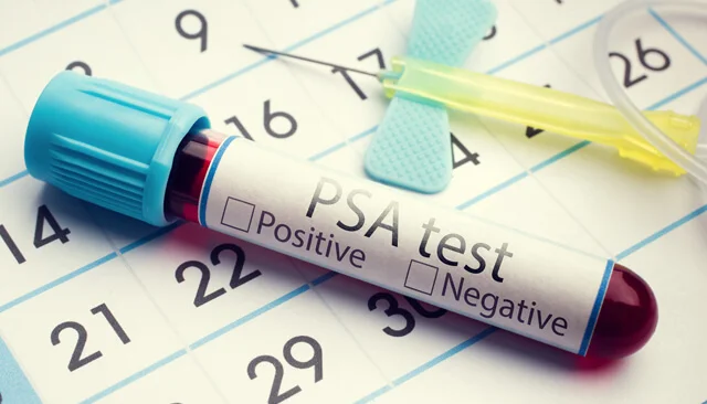
An elevated PSA level can be caused not only by prostate cancer but also by prostatitis (inflammation of the prostate) or benign prostatic hyperplasia (BPH). Therefore, if PSA is above 2 ng/mL, additional diagnostic evaluations are necessary.
In recent years, multiparametric Magnetic Resonance Imaging (mpMRI) of the prostate has been recommended in such cases by Western guidelines.
What Are the Disadvantages of Prostate mpMRI?
-
It is relatively expensive.
-
Accurate interpretation of mpMRI results requires a radiologist with specific expertise in prostate cancer.
-
It requires the patient to lie still in a closed space for an extended period of time.
-
It is not a complete method for performing a biopsy—real-time (“live”) prostate biopsy cannot be done directly with MRI. Therefore, fusion biopsy becomes necessary.
What Does “Fusion” Biopsy Mean (in the Era of Micro-Echoscopia)?
The word “fusion” in Georgian means combining. A prostate biopsy refers to the process of taking small tissue “samples” from the prostate using a special needle. In a “fusion” biopsy, two research methods are combined: Magnetic Resonance Imaging (MRI) and traditional ultrasound (echoscopy). The combination of these two techniques allows for the collection of tissue from suspicious areas of the prostate identified in the MRI. The fusion, or “fusion,” occurs using a special computer program. The suspicious areas on the MRI disc are projected onto the ultrasound image so that the biopsy can be performed with traditional ultrasound guidance from those specific suspicious regions.
Is There Still a Place for Traditional Systemic Biopsy with Traditional Ultrasound Guidance in the Era of Micro-Echoscopia and “Fusion” Biopsy?
Prostate systemic biopsy with traditional ultrasound guidance is now considered an outdated method because traditional ultrasound is “blind.” Traditional ultrasound misses prostate cancer in approximately 40% of cases. Thus, using traditional ultrasound for targeting could cause the needle to miss the cancer, and to make an accurate diagnosis, the prostate needs to be “shot” from multiple locations. However, if the tumor is small, it might still evade the needle, leading to an incorrect diagnosis. Therefore, traditional prostate systemic biopsy with ultrasound guidance is inaccurate and outdated, and it no longer has a place in modern diagnostics.
In the era of micro-ecoscopia, fusion biopsy, which combines MRI and traditional ultrasound, was considered the most advanced method.
What Are the Disadvantages of “Fusion” Biopsy?
-
Direct MRI-guided prostate biopsy is technically impossible, while traditional ultrasound is “blind.” Therefore, the “fusion” biopsy is an attempt to combine two imperfect methods into one perfect method, but this combination is artificially done electronically through a special computer program and not in a live, “real-time” mode.
-
It is expensive, as it requires both MRI and traditional ultrasound.
-
It takes a lot of time, and it is very inconvenient for the patient because they first need to schedule an MRI, undergo the MRI procedure, then schedule the biopsy, and finally undergo the biopsy.
What Is the Solution to These Challenges?
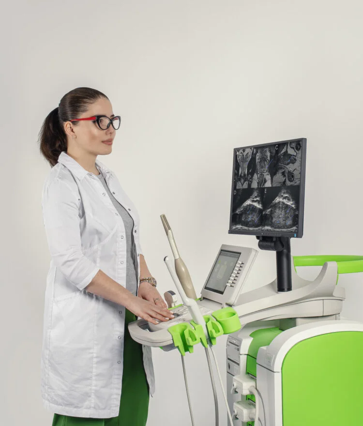
The solution is microscopic ultrasound, or micro-ecoscopia!
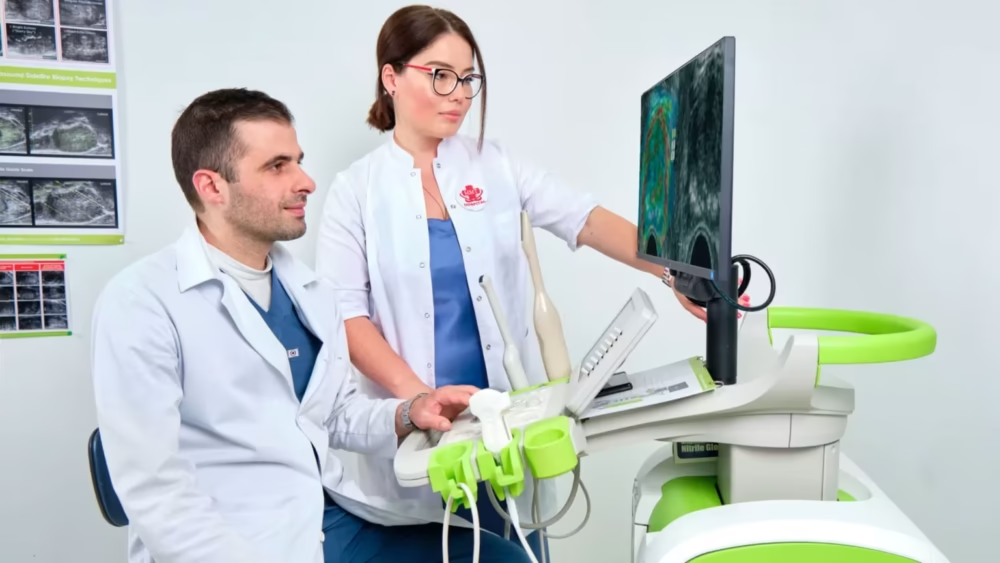
What Are the Advantages of Micro-Echoscopia Compared to MRI and Traditional Ultrasound?
-
It is an ultra-modern technique with no precedent or analog in the world.
-
Prostate micro-echoscopia provides an image that is 300% more detailed than traditional ultrasound, allowing for the visualization of even the tiniest structures, as small as the diameter of a hair.
-
The diagnostic efficacy of micro-ecoscopia for detecting prostate cancer is comparable to, or even better than, MRI.
-
Micro-echoscopia is significantly cheaper than MRI.
-
Micro-echoscopia is faster than MRI.
-
Micro-echoscopia is performed by the urologist or in urology clinics that have more experience in prostate cancer diagnostics, ensuring that high-quality, accurate image interpretation is guaranteed.
-
Micro-echoscopia has eliminated the need for MRI and traditional ultrasound fusion (“fusion” biopsy). However, if desired, the highest standard “fusion” can still be performed, which combines MRI and microscopic ultrasound.
Why Has Micro-Echoscopia Eliminated the Need for “Fusion” Biopsy?
-
Micro-echoscopia detects prostate cancer so well that MRI is no longer necessary.
-
Micro-echoscopia allows for “live” targeted, or guided, biopsies. In other words, once micro-echoscopia has “seen” the target, the biopsy can be performed immediately at the identified site, without the need for MRI or a special computer program to combine MRI and traditional ultrasound.
-
Biopsies performed with micro-echoscopia are more affordable, as they no longer require MRI.
-
Micro-echoscopia saves the patient’s time! There is no need to schedule an MRI, undergo the MRI, then schedule the biopsy, and so on. Everything happens quickly in one place with one visit.
If the patient desires, MRI and micro-echoscopia fusion biopsy can still be performed. There is no higher standard for prostate cancer diagnosis worldwide!
Conclusion:
What is modern prostate cancer screening with cutting-edge technology and the latest Western recommendations?
-
Essential test: New PSA blood analysis norm — 2 ng/ml.
-
Essential test: Prostate micro-echoscopia.
-
If necessary: Targeted or guided biopsy under the control of micro-echoscopia.
-
If the patient desires: “Fusion” biopsy combining MRI and micro-echoscopia.
Karazanashvilis Robotic Center is the only clinic in the region (Georgia, Turkey, Russia, Armenia, Azerbaijan) that offers modern prostate cancer screening using micro-echoscopia! For this, we use the latest Canadian-made ExactVu equipment.