Computed Tomography (CT) is a state-of-the-art diagnostic technology used in medicine that helps doctors study the internal structures of the human body in detail. A CT scanner creates cross-sectional images of internal organs and tissues.
What information does computed tomography provide to the doctor and patient?
CT helps doctors obtain detailed information about internal organs, tissues, and bones. It allows for the diagnosis of a wide range of diseases, the assessment of damage levels, and the evaluation of treatment effectiveness. The Karazanashvili Robotic Center focuses on patients with urological problems and oncology, so the main reasons for our patients’ CT visits are:
- Study of abdominal and pelvic organs (diagnosis of inflammatory processes, appendicitis, pancreatitis).
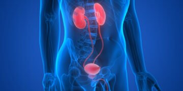
- Detection of stones in the kidneys and gallbladder.
- Identification of issues in the intestines, liver, spleen, or pancreas.
- Detection of tumors (diagnosis of malignant and benign growths, assessment of tumor size, shape, and location, detection of metastases in other organs).
Patients often come for treatment monitoring, which involves:
- Monitoring the effectiveness of chemotherapy or radiation therapy.
- Evaluating the results of surgical interventions.
- Monitoring changes in the body’s condition over time.
CT also allows for research in cases where other methods, such as X-rays or ultrasound, are insufficiently informative.
Device Data and Advantages
At the Karazanashvili Robotic Center, you’ll find the Toshiba AQUILION PRIME 302a 160 slices – this is a third-generation CT scanner with 160 slices. This gives it several advantages over other CT scanners, including:
- High resolution, providing precision of 0.1-0.3 mm.
- Scanning 2-3 times faster, as the device scans 160 slices simultaneously in one rotation, which also reduces the patient’s radiation dose by 75%.
- Many patients come with claustrophobia issues, but this model features a wide tunnel, making the scanning process as comfortable as possible.
How is the CT procedure performed?
The CT procedure is usually simple and quick. Patients typically come upon a doctor’s referral, allowing the radiologic technologist to select the ideal scanning protocol for the specific request.
Sometimes, a contrast agent is used to better visualize the organs, which is injected intravenously or orally.
During the procedure, you will be asked to lie on a special table that moves into the CT scanner. You must remain still, as movement can degrade image quality. The medical staff will position the scanner so it focuses on the specific area of your body. Only the table and the CT scanner will remain in the room, while doctors will monitor the process from an adjacent room through glass. The machine will start, and X-ray beams will pass through your body. You may hear a faint noise or clicking sounds. Occasionally, you will be instructed to “remain still” or “hold your breath” (if a chest or abdominal scan is being done).
Next, the images will be sent for processing. The radiologist (a doctor specializing in interpreting CT images) will study them and send the results to your treating physician. Results typically take anywhere from a few hours to a few days.
Procedure Duration:
- Regular CT: 5-10 minutes.
- CT with contrast: 20-40 minutes, including the administration of contrast material.
Please note, CT scans are based on X-ray radiation and should be prescribed by your treating physician, who will assess all the risks and benefits of the scan. CT is not a comprehensive examination, and your physician may prescribe additional tests and laboratory analyses.